Bisferiens Pulse
| Pulsus bisferiens | |
|---|---|
| Other names | Bisferious pulse, biphasic pulse |
| Specialty | Cardiology |

تاکیکاردی سینوسی (به انگلیسی: Sinus tachycardia); تاکیکاردی بطنی (به انگلیسی: Ventricular tachycardia): ضربآهنگ تند نابهنجار بیش از ۱۵۰ ضربان در دقیقه که از بطن سرچشمه میگیرد و عموماً با گسستگی دهلیزی ـ بطنی (به انگلیسی. Time is important factor in the studiesVideo is made very short & all information is given in short video.Video covers all aspects of the topic Video is made.
Bigeminal Pulse
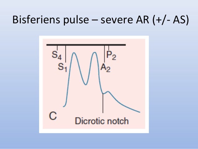
Pulsus bisferiens, also known as biphasic pulse, is an aortic waveform with two peaks per cardiac cycle, a small one followed by a strong and broad one.[1] It is a sign of problems with the aorta, including aortic stenosis and aortic regurgitation, as well as hypertrophic cardiomyopathy causing subaortic stenosis.[1]
Pathogenesis[edit]
In hypertrophic cardiomyopathy, there is narrowing of the left ventricular outflow tract (LVOT) due to hypertrophy of the interventricular septum. During systole, the narrowing of the LVOT creates a more negative pressure due to the Venturi effect and sucks in the anterior mitral valve leaflet. This creates a transient occlusion of the LVOT, causing a midsystolic dip in the aortic waveform. Towards the end of systole, the ventricle is able to overcome the obstruction to cause the second rise in the aortic waveform.[2]
In severe aortic regurgitation, additional blood reenters the left ventricle during diastole. This added volume of blood must be pumped out during ventricular systole. The rapid flow of blood during systole is thought to draw the walls of the aorta together due to the Venturi effect, temporarily decreasing blood flow during midsystole.[2]
A recent paper theorized that an alternative explanation for pulsus bisferiens may be due to a forward moving suction wave occurring during mid-systole.[3]
References[edit]
- ^ abRiojas CM, Dodge A, Gallo DR, White PW (January 2016). 'Aortic Dissection as a Cause of Pulsus Bisferiens: A Case Report and Review'. Annals of Vascular Surgery. 30: 305.e1–5. doi:10.1016/j.avsg.2015.07.026. PMID26520426.
- ^ abMcGee SR (2018). 'Chapter 15: Pulse Rate and Contour'. Evidence-based physical diagnosis (Fourth ed.). Philadelphia, PA: Elsevier. pp. 95–108. ISBN978-0-323-50871-1. OCLC959371826.
- ^Chirinos JA, Akers SR, Vierendeels JA, Segers P (March 2018). 'A Unified Mechanism for the Water Hammer Pulse and Pulsus Bisferiens in Severe Aortic Regurgitation: Insights from Wave Intensity Analysis'. Artery Research. 21: 9–12. doi:10.1016/j.artres.2017.12.002. PMC5863934. PMID29576810.
External links[edit]
| Classification |
|
|---|
Pathogenesis
In hypertrophic cardiomyopathy, there is narrowing of the left ventricular outflow tract (LVOT) due to hypertrophy of the interventricular septum. During systole, the narrowing of the LVOT creates a more negative pressure due to the Venturi effect and sucks in the anterior mitral valve leaflet. This creates a transient occlusion of the LVOT, causing a midsystolic dip in the aortic waveform. Towards the end of systole, the ventricle is able to overcome the obstruction to cause the second rise in the aortic waveform.[2]
Bisferiens Pulse Is Characteristically Found In
In severe aortic regurgitation, additional blood reenters the left ventricle during diastole. This added volume of blood must be pumped out during ventricular systole. The rapid flow of blood during systole is thought to draw the walls of the aorta together due to the Venturi effect, temporarily decreasing blood flow during midsystole.[2]
Pulsus Differens
A recent paper theorized that an alternative explanation for pulsus bisferiens may be due to a forward moving suction wave occurring during mid-systole.[3]
